
831
PerssonAP, etal. Heart 2024;110:831–837. doi:10.1136/heartjnl-2023-323681
Original research
Reference ranges for ambulatory heart rate
measurements in a middle- agedpopulation
Anders Paul Persson ,
1,2
Alexandra Måneheim,
1,2
Johan Economou Lundeberg,
1,3
Artur Fedorowski ,
4,5
Jeff S Healey,
6,7
Johan Sundström ,
8,9
Gunnar Engström ,
1
Linda S B Johnson
1,6
Arrhythmias and sudden death
To cite: PerssonAP,
MåneheimA, Economou
LundebergJ, etal. Heart
2024;110:831–837.
► Additional supplemental
material is published online
only. To view, please visit the
journal online (https:// doi.
org/ 10. 1136/ heartjnl- 2023-
323681).
1
Department of Clinical
Sciences, Lund University,
Malmö, Sweden
2
Department of Clinical
Physiology, Skånes
universitetssjukhus Malmö,
Malmö, Sweden
3
Department of Clinical
Physiology, Skånes
universitetssjukhus Lund, Lund,
Sweden
4
Department of Clinical
Sciences, Lund University Faculty
of Medicine, Malmö, Sweden
5
Department of Medicine,
Karolinska Institute, Solna,
Sweden
6
Population Health Research
Institute, Hamilton, Ontario,
Canada
7
Department of Medicine,
McMaster University, Hamilton,
Ontario, Canada
8
Department of Medical
Sciences, Uppsala University,
Uppsala, Sweden
9
The George Institute for Global
Health, Newtown, New South
Wales, Australia
Correspondence to
Dr Anders Paul Persson,
Department of Clinical Sciences,
Lund University, Malmö 205 02,
Sweden;
anders_ p. persson@ med. lu. se
Received 9 November 2023
Accepted 10 March 2024
Published Online First
5April2024
© Author(s) (or their
employer(s)) 2024. Re- use
permitted under CC BY.
Published by BMJ.
ABSTRACT
Background Elevated heart rate (HR) predicts
cardiovascular disease and mortality, but there are no
established normal limits for ambulatory HR. We used
data from the Swedish CArdioPulmonary Imaging
Study to determine reference ranges for ambulatory HR
in a middle- aged population. We also studied clinical
correlates of ambulatory HR.
Methods A 24- hour ECG was registered in 5809 atrial
fibrillation- free individuals, aged 50–65 years. A healthy
subset (n=3942) was used to establish reference values
(excluding persons with beta- blockers, cardiovascular
disease, hypertension, heart failure, anaemia, diabetes,
sleep apnoea or chronic obstructive pulmonary disease).
Minimum HR was defined as the lowest 1- minute HR.
Reference ranges are reported as means±SDs and 2.5th–
97.5th percentiles. Clinical correlates of ambulatory HR
were analysed with multivariable linear regression.
Results The average mean and minimum HRs were
73±9 and 48±7 beats per minute (bpm) in men and
76±8 and 51±7 bpm in women; the reference range
for mean ambulatory HR was 57–90 bpm in men and
61–92 bpm in women. Average daytime and night- time
HRs are also reported. Clinical correlates, including age,
sex, height, body mass index, physical activity, smoking,
alcohol intake, diabetes, hypertension, haemoglobin level,
use of beta- blockers, estimated glomerular filtration rate,
per cent of predicted forced expiratory volume in 1 s and
coronary artery calcium score, explained <15% of the
interindividual differences in HR.
Conclusion Ambulatory HR varies widely in healthy
middle- aged individuals, a finding with relevance for
the management of patients with a perception of
tachycardia. Differences in ambulatory HR between
individuals are largely independent of common clinical
correlates.
INTRODUCTION
High resting heart rates have been linked to cardio-
vascular, cancer and all- cause death.
1
The finding
has been consistent in population- based studies,
2–4
as well as in studies of individuals with hyperten-
sion,
5
diabetes,
6
chronic obstructive pulmonary
disease,
7
cardiovascular disease
8
and heart failure.
9
Low heart rate, on the other hand, is associated with
atrial fibrillation.
10–12
Regardless of whether these
associations imply a causal relation between heart
rate and outcomes or not, heart rate measurements
can be useful for disease prediction. Ambulatory
heart rates can be inexpensively, reliably and non-
invasively measured, and ambulatory ECG moni-
toring is frequently performed in clinical practice.
Furthermore, patients with post- COVID- 19 condi-
tion frequently report inappropriately elevated
resting heart rates,
13 14
a symptom which cannot be
put in context without knowledge of normal limits.
Despite this, studies that report reference ranges
and or predictors of heart rate at 24- hour ECG
(24hECG) are lacking.
We aimed to describe reference ranges for, and
predictors of ambulatory heart rate measured with
24hECG in a middle- aged general population
sample.
METHODS
Study sample
The population- based Swedish CArdioPulmonary
Imaging Study (SCAPIS) cohort included 30 154
participants aged 50–65 years old recruited from
the general population, in six municipal centres
containing university hospitals in Sweden (Gothen-
burg, Linköping, Malmö, Stockholm, Uppsala and
Umeå). Examination with 24hECG was part of the
protocol at the Malmö (6235 participants) and
Uppsala (5038 participants) sites. All participants
examined in Malmö between September 2016 and
2018 as well as all participants in Uppsala were
asked to contribute a 24hECG registration, of
which 1288 (Malmö) and 5004 (Uppsala) accepted.
WHAT IS ALREADY KNOWN ON THIS TOPIC
⇒ Ambulatory heart rate varies widely and is
only explained by clinical correlates to a minor
degree.
WHAT THIS STUDY ADDS
⇒ This study presents reference ranges of
ambulatory heart rate in a middle- aged
population.
HOW THIS STUDY MIGHT AFFECT RESEARCH,
PRACTICE OR POLICY
⇒ These normal limits for heart rate can be used
for the interpretation of ambulatory ECGs, for
instance, to determine whether patients with
subjective perception of tachycardia have
abnormally elevated heart rates.
on August 29, 2024 by guest. Protected by copyright.http://heart.bmj.com/Heart: first published as 10.1136/heartjnl-2023-323681 on 5 April 2024. Downloaded from

832
PerssonAP, etal. Heart 2024;110:831–837. doi:10.1136/heartjnl-2023-323681
Arrhythmias and sudden death
After excluding individuals with a previous diagnosis of atrial
fibrillation (n=112), atrial fibrillation during the ambula-
tory ECG recording (n=35), registration duration <16 hours
(n=77) or insufficient 24hECG registration quality (n=258),
and one participant with an implausible mean heart rate (155
beats per minute (bpm)), 5809 participants constituted the final
study population (figure 1). Reference ranges for heart rate
were reported in a healthy population subset after exclusion
Figure 1 Derivation of study population. 24hECG, 24- hour ECG; SCAPIS, Swedish CArdioPulmonary Imaging Study.
Table 1 Sample characteristics
All (N=5809) Women Men Healthy reference sample (n=3942)
Age, mean (range) 58 (50–65) 58 (50–65) 58 (50–65) 57 (50–65)
Women, % 53 – – 54
Height, cm (SD) 172 (9.7) 166 (6.5) 179 (7.0) 172 (9.7)
BMI, %
<25 35 42 27 40
25–30 44 38 51 44
>30 21 20 22 16
Smoking, %
Never 56 53 59 58
Former 34 37 30 32
Current 11 11 11 10
Alcohol intake units/week, median (IQR) 2.6 (1.1–4.1) 1.1 (0.4–3.8) 2.6 (1.1–6.0) 2.6 (1.1–4.1)
Physical activity, %
Low 57 60 54 54
High 43 40 46 46
Diabetes, % 25 7 11 –
Hypertension, % 21 20 22 –
Use of oral beta- blockers, % 5,8 6.1 5.5 –
FEV
1
%predicted (SD) 109 (15) 109 (15) 108 (14) 110 (14)
Coronary artery calcium score, %
0 61 74 46 67
1–99 27 21 35 25
≥100 11 5 19 8
eGFR, mL/min/1.73 m
2
(IQR) 88 (79–96) 88 (78–96) 89 (80–96) 88 (79–96)
Haemoglobin, g/L (SD) 142 (12) 135 (9.1) 149 (9.4) 142 (11)
Sleep apnoea, % 3.7 2.6 4.9 –
Coronary artery disease, % 2.0 1.1 3.0 –
Heart failure, % 0.3 0.3 0.4 –
BMI, body mass index; eGFR, estimated glomerular filtration rate; FEV
1
%predicted, per cent of predicted forced expiratory volume during 1 s.
on August 29, 2024 by guest. Protected by copyright.http://heart.bmj.com/Heart: first published as 10.1136/heartjnl-2023-323681 on 5 April 2024. Downloaded from

833
PerssonAP, etal. Heart 2024;110:831–837. doi:10.1136/heartjnl-2023-323681
Arrhythmias and sudden death
of participants with known prevalent cardiovascular diseases,
hypertension, heart failure, anaemia, diabetes, obstructive sleep
apnoea, chronic obstructive pulmonary disease, and individuals
using beta- blockers, resulting in a healthy reference cohort of
3942 subjects. Neither the study participants nor the public
were involved in the design, conduct, reporting or dissemination
plans of this study.
Data collection
24hECGs were recorded using CardioSpy equipment (Labtech,
Debrecen, Hungary), with X, Y, Z coupling and sampling
frequency of 256 Hz, and measures of heart rate were derived
from the CardioSpy ECG analysis software. Cases of atrial fibril-
lation during 24hECG were detected using a Food and Drug
Association and CE- approved artificial intelligence algorithm
(MEDICALgorithmics, Warsaw, Poland). The measures of heart
rate included in this study were mean heart rate during the
entire recording, minimum heart rate during 1 min, and mean
day (06:00–22:00) and night- time (22:00–06:00) heart rates.
Nightly dip in heart rate was calculated as daytime heart rate
minus night- time heart rate. Maximum heart rate during 1 min
was not studied in detail due to its dependence on maximum
exertion but percentiles are presented in online supplemental
table 1.
Each participant completed an extensive health and lifestyle
questionnaire from which data concerning physical activity,
smoking and alcohol use were obtained, as was the prevalence
of known prevalent diseases and drug treatment. A capillary
glucose sample was collected, and if elevated (≥7 mmol/L), a
repeat measurement was performed on another day to diagnose
diabetes.
Height was measured to the nearest centimetre and weight
was measured on a digital scale in light indoor clothing without
shoes. Body mass index (BMI) was calculated as kg/m
2
and
stratified at 25 kg/m
2
, 25–30 kg/m
2
and >30 kg/m
2
. Dynamic
spirometry (Jaeger MasterScreen PFT; Carefusion, Hoech-
berg, Germany) was performed 15 min after bronchodilation
using 400 µg salbutamol with subjects in the sitting position
and wearing a nose clip, and forced expiratory volume during
1 s (FEV
1
) was measured according to the American Thoracic
Society and European Respiratory Society standards.
15 16
The
Hedenström formula was used to calculate per cent of predicted
FEV
1
(FEV
1
%predicted).
17 18
Smoking status was categorised as
current, former or never smoker. Reported leisure time physical
activity was categorised as low (mostly sedentary or some light
physical activity) or high (physical activity with moderate to
strenuous intensity at least 2 hours per week). Alcohol intake in
units per week was calculated by multiplying average times per
week of drinking during the last year with the typical intake on
a day of drinking. Alcohol intake was then stratified into two
groups (above or below the median intake of 2.6 units per week).
A fasting venous blood sample was retrieved and haemoglobin
and creatinine concentrations were measured using standard
laboratory procedures at the Uppsala and Malmö University
Hospitals. Estimated glomerular filtration rate (eGFR) was
calculated using the creatinine- based Chronic Kidney Disease
Epidemiology Collaboration formula.
19
CT was performed in all participants using Siemens Defini-
tion Flash 2×128 slice, stellar detector, 4D‐Care dose, Care‐kV
and sinogram‐affirmed iterative reconstruction (Forchheim,
Germany). Coronary artery calcium score was calculated using
the Agatston score
20
and stratified into three groups: 0, 1–99
and ≥100.
Statistical analyses
Reference ranges for heart rates are reported as means (±SD
and percentiles) and presented in histograms and sex- specific
cumulative distribution function plots. The reported percen-
tiles were taken directly from the observed distribution and not
derived from the SD. The sex- specific cumulative distribution
function plots for average day and night- time heart rate are
presented in online supplemental figure 1. Multivariable linear
regression models were used to analyse the association between
heart rate and a prespecified set of predictors including age,
sex, height, BMI, physical activity, smoking, alcohol intake,
diabetes, hypertension, haemoglobin level, use of beta- blockers,
eGFR, FEV
1
%predicted and coronary artery calcium score. All
continuous parameters were assessed visually in histograms for
normality. Collinearity was ruled out using the variance inflation
factor, using a cut- off of 2.5. Model diagnostics were performed
using plots of residuals and fitted values and histograms of
the residuals. No violations of the assumptions were observed
(online supplemental figures 2–5). Linear regression robust to
heteroskedasticity was also performed for mean and minimum
heart rate with virtually unchanged results (online supplemental
table 3).
All statistical analyses were performed using Stata V.15.1
(StataCorp, College Station, Texas, USA).
Table 2 Reference ranges for measures of ambulatory heart rate
Mean heart rate (beats/min) Minimum heart rate* (beats/min) Average daytime heart rate (beats/min) Average night- time heart rate (beats/min)
Men Women Men Women Men Women Men Women
Healthy individuals (1825 men and 2117 women)†
Mean (SD) 73 (9) 76 (8) 48 (7) 51 (7) 78 (9) 81 (8) 63 (9) 65 (8)
Percentiles
2.5 57 61 36 39 60 65 48 51
5 59 63 38 40 63 67 50 53
25 67 71 44 46 71 75 57 60
50 73 76 48 51 78 81 63 65
75 79 81 53 55 84 86 68 71
95 87 89 60 62 93 94 79 80
97.5 90 92 63 65 96 97 83 83
*Lowest average heart rate during 1 min.
†Based on 3942 (2117 women) healthy individuals, excluding individuals with new or known diabetes (490) or known prevalent hypertension (n=995), sleep apnoea (n=111), coronary
artery disease (n=39), chronic obstructive pulmonary disease (n=32), heart failure (n=6) or anaemia (haemoglobin <120 g/L in women and <130 g/L in men) (n=126, missing 15) and
individuals using beta- blockers (54).
on August 29, 2024 by guest. Protected by copyright.http://heart.bmj.com/Heart: first published as 10.1136/heartjnl-2023-323681 on 5 April 2024. Downloaded from
834
PerssonAP, etal. Heart 2024;110:831–837. doi:10.1136/heartjnl-2023-323681
Arrhythmias and sudden death
RESULTS
The age of study participants was evenly distributed between
50 and 65 years of age and 52% were women. While 22% had
hypertension, heart failure was rare (0.3%). Study sample char-
acteristics are presented in more detail in table 1.
Reference ranges for heart rate
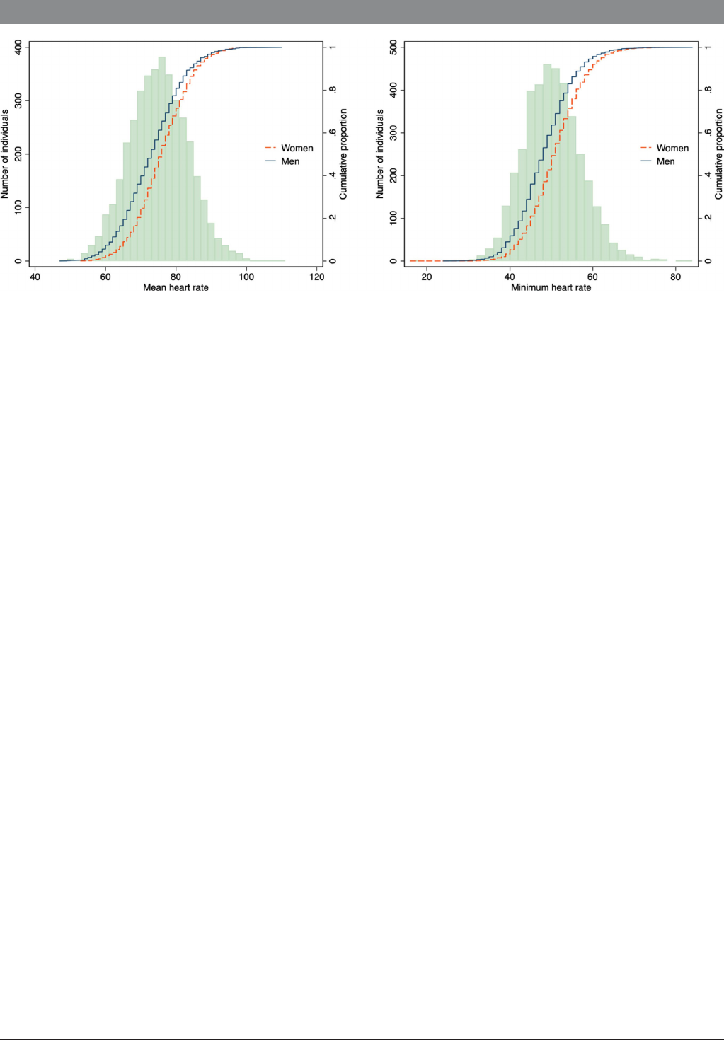
A healthy population subset was used to determine reference
ranges for heart rate measures. The mean heart rate was 73
(±9) bpm on average for men and 76 (±8) bpm on average
for women, and the minimum heart rate was 48 (±7) bpm on
average for men and 51 (±7) bpm on average for women.
The range of heart rate was wide; the 2.5th–97.5th percentile
of mean heart was 57–90 bpm in men and 61–92 bpm in women
(table 2 and figure 2). Reference heart rates (only including
healthy individuals) did not differ substantially from heart rates
in the entire study population (online supplemental table 2). The
average nightly dip in heart rate was 15 bpm in men and 16 bpm
in women. Heart rate parameters are reported in detail for both
men and women in table 2.
Clinical predictors of heart rate
Clinical predictors of heart rate are calculated in the full study
sample and presented in table 3. We included 14 clinical variables
in the multivariable linear regression, and 11 (sex, height, BMI,
smoking, high physical activity, alcohol intake, diabetes, using
oral beta- blockers, FEV
1
%predicted, eGFR and haemoglobin)
were statistically significantly associated with mean heart rate
(table 3). The associations were overall rather weak, however.
For example, the mean heart rate was on average only 4.5 bpm
lower in beta- blocker users and 2.7 bpm higher in smokers,
after multivariable adjustment (table 3). In keeping with this,
the variation in heart rate was explained by the clinical vari-
ables included in the multivariable model only to a small degree;
adjusted R
2
and unadjusted R
2
in the multivariable model were
<15% and <16%, respectively, for all the studied measures of
heart rate. In sex- stratified multivariable models, the adjusted R
2
for mean heart rate was 16% in men and 10% in women. Within
the age range of this study, heart rate was virtually unchanged
with age.
DISCUSSION
This is the largest population- based study of heart rate at ambu-
latory ECG to date, and the first to provide reference ranges for
measures of heart rate at 24hECG in a middle- aged population.
We found the interindividual differences in heart rate to be large
and largely independent of clinical risk factors for cardiovascular
disease.
Previous studies have shown that there is prognostic value in
ambulatory heart rate measurements,
21
but since reference ranges
have been lacking, it has not been clear how ambulatory heart
rate measurements could be used in clinical practice. This matter
has become even more relevant in the context of the COVID- 19
pandemic and the emergence of post- COVID- 19 syndromes,
where a feeling of increased heart rate is often reported, but
objective guidelines as to what constitutes elevated heart rate and
pre- COVID- 19 references for ambulatory heart rate have been
lacking. For instance, although inappropriate sinus tachycardia is
typically diagnosed when average heart rate on 24hECG moni-
toring exceeds 90 bpm,
22
population- based studies supporting
this diagnostic threshold are limited, and definitions in current
literature are inconsistent.
23
Data in this study were collected
before the pandemic, ensuring that the study population is free
from patients suffering from post- COVID- 19 condition.
We found less than one- sixth of the differences in ambula-
tory heart rate measurements to be explained by risk factors for
cardiovascular disease, so most of the interindividual differences
in heart rate remain unexplained. Even so, markers for poor
health (such as inactivity, smoking, higher BMI and lower lung
function) were associated with a higher heart rate. Somewhat
surprisingly, worse kidney function (ie, lower eGFR) was associ-
ated with a lower mean and minimum heart rate. One previous
study has shown that lower eGFR is associated with chrono-
tropic incompetence in patients with heart failure with preserved
ejection fraction.
24
We also found lower haemoglobin levels to be associated
with lower heart rate, which might seem contraintuitive, since
lower haemoglobin physiologically should lead to a compensa-
tory increase in heart rate. This association could perhaps be
explained by unmeasured confounding (for example, by dehy-
dration, which can cause both an increase in heart rate and a
higher level of haemoglobin), an indication that there could be
other unknown determinants of heart rate that our relatively
extensive model does not include.
Causal pathways between heart rate and cardiovascular
disease have been suggested,
25–27
but in most studies, heart rate
is associated with a similar risk increase for all- cause mortality,
cancer mortality and cardiovascular mortality,
1
which could
Figure 2 Heart rates in the healthy reference sample, histogram and sex- specific cumulative distribution curves.
on August 29, 2024 by guest. Protected by copyright.http://heart.bmj.com/Heart: first published as 10.1136/heartjnl-2023-323681 on 5 April 2024. Downloaded from

835
PerssonAP, etal. Heart 2024;110:831–837. doi:10.1136/heartjnl-2023-323681
Arrhythmias and sudden death
indicate that higher heart rates are not causally related to cardio-
vascular disease, but rather markers of poor health. On the other
hand, a genome- wide association study has identified heart rate-
associated genes which explain 2.5% of the differences in resting
heart rate and shown these genes to be associated with increased
all- cause mortality,
28
implying that a causal component to the
association between heart rate and mortality may exist. Heart
rate only slightly increases the risk of stroke,
6
which is likely
due to the fact that the association between low heart rate and
atrial fibrillation is in the opposite direction from the association
between heart rates and cardiovascular disease
12
—two major
stroke risk factors with different mechanisms.
Limitations
We have used a large population- based study with extensive infor-
mation on comorbidities and lifestyle to describe reference ranges
and predictors for ambulatory measures of heart rate in the general
population. Some limitations exist that need to be considered. The
cross- sectional nature of this study does not allow any estimation
Table 3 Multivariable linear regression models for ambulatory heart rate measures
N=5021
Mean heart rate, beats/min
(95% CI)
Minimum heart rate,
beats/min (95% CI)
Average daytime heart
rate, beats/min (95% CI)
Average night- time heart
rate, beats/min (95% CI)
Beta (95% CI) t ratio* Beta (95% CI) t ratio* Beta (95% CI) t ratio* Beta (95% CI) t ratio*
Age (per 1 year) −0.0
(−0.1, 0.0)
−1.4 0.1
(0.0, 0.1)
3.3 −0.0
(−0.1, 0.0)
−1.4 −0.0
(−0.1, 0.0)
−0.6
Men (vs women) −2.8
(−3.5, −2.0)
−7.3 −2.2
(−2.9, −1.6)
−7.1 −3.2
(−4.0, −2.4)
−7.7 −2.0
(−2.8, −1.3)
−5.2
Height (per 10 cm) −0.8
(−1.2, −0.5)
−4.9 −0.8
(−1.0, −0.5)
−5.2 −0.8
(−1.2, −0.5)
−4.4 −0.9
(−1.2, −0.6)
−5.1
Body mass index, kg/m
2
<25 Ref Ref Ref Ref Ref Ref Ref Ref
25–30 0.9
(0.4, 1.4)
3.5 0.8
(0.4, 1.3)
3.8 0.6
(0.1, 1.2)
2.3 1.4
(0.9, 2.0)
5.4
>30 1.9
(1.2, 2.6)
5.6 1.4
(0.9, 2.0)
5.1 1.4
(0.7, 2.1)
3.9 2.9
(2.3, 3.6)
8.5
Smoking
Never Ref Ref Ref Ref Ref Ref Ref Ref
Former 1.2
(0.7, 1.7)
4.9 0.9
(0.5, 1.3)
4.3 1.1
(0.6, 1.7)
4.2 1.3
(0.8, 1.9)
5.2
Current 2.7
(1.9, 3.4)
6.7 2.2
(1.6, 2.9)
6.8 2.2
(1.3, 3.0)
5.0 3.7
(2.9, 4.5)
9.2
High physical activity (vs low) −3.6
(−4.1, −3.1)
−15.1 −2.7
(−3.1, −2.3)
−13.5 −3.7
(−4.2, −3.2)
−14.4 −3.3
(−3.8, −2.9)
−13.7
Alcohol intake, above median (vs below median) 0.7
(0.2, 1.1)
3.0 0.8
(0.4, 1.2)
4.1 0.5
(0.0, 1.0)
2.1 1.0
(0.6, 1.5)
4.3
Diabetes (yes vs no) 2.0
(1.1, 2.8)
4.5 2.1
(1.4, 2.8)
5.8 1.7
(0.8, 2.6)
3.6 2.5
(1.6, 3.4)
5.5
Hypertension (yes vs no) 0.4
(−0.1, 1.0)
1.5 0.1
(−0.4, 0.6)
0.4 0.5
(−0.1, 1.2)
1.6 0.3
(−0.3, 0.9)
0.8
Using oral beta- blockers (yes vs no) −4.5
(−5.5, −3.4)
−8.5 −0.9
(−1.8, −0.1)
−2.2 −5.7
(−6.8, −4.5)
−9.9 −1.8
(−2.8, −0.7)
−3.3
FEV
1
%predicted, per 10% increase −0.3
(−0.4, −0.1)
−3.6 −0.3
(−0.5, −0.2)
−4.8 −0.2
(−0.4, 0.0)
−2.4 −0.4
(−0.6, −0.3)
−5.3
Coronary artery calcium score
0 Ref Ref Ref Ref Ref Ref Ref Ref
1–99 0.4
(−0.1, 1.0)
1.6 0.4
(−0.1, 0.8)
1.6 0.4
(−0.2, 1.0)
1.3 0.5
(0.0, 1.1)
1.8
≥100 0.6
(−0.2, 1.4)
1.5 0.5
(−0.1, 1.2)
1.6 0.5
(−0.4, 1.3)
1.1 0.8
(0.0, 1.6)
2.0
eGFR (per 10 mL/min/1.73 m
2
) 0.6
(0.4, 0.8)
6.1 0.4
(0.3, 0.6)
5.0 0.7
(0.5, 0.9)
6.2 0.5
(0.3, 0.7)
5.1
Haemoglobin (per 10 g/L increase) 0.7
(0.5, 1.0)
5.7 0.3
(0.1, 0.5)
2.5 0.8
(0.6, 1.1)
6.1 0.4
(0.2, 0.7)
3.5
Constant† 76.1
(75.4, 76.8)
49.9
(49.3, 50.5)
81.5
(80.7, 82.3)
64.4
(63.7, 65.2)
Unadjusted R
2
0.149 0.143 0.133 0.151
Adjusted R
2
0.146 0.140 0.130 0.148
Bold numbers indicate statistical significance (p<0.05).
*Larger t ratios (both positive and negative) imply larger impacts on heart rate.
†The constant is what is expected for an individual with values that have been set as reference as stated in the table and age 50 years old, the mean of the continuous variables
height (172 cm), eGFR (89 mL/min/1.73 m
2
), haemoglobin (142 g/L) and FEV
1
%predicted (109%).
eGFR, estimated glomerular filtration rate; FEV
1
%predicted, per cent of predicted forced expiratory volume during 1 s.
on August 29, 2024 by guest. Protected by copyright.http://heart.bmj.com/Heart: first published as 10.1136/heartjnl-2023-323681 on 5 April 2024. Downloaded from

836
PerssonAP, etal. Heart 2024;110:831–837. doi:10.1136/heartjnl-2023-323681
Arrhythmias and sudden death
of the predictive value of different ambulatory heart rate ranges,
and though we found large interindividual differences in heart rates
even among otherwise healthy individuals, we are not able to study
whether ambulatory heart rates predict or cause incident disease.
There are also some potential sources of measurement error. We
adjusted for habitual leisure time physical activity, but mean heart rate
is reasonably also affected by activity during the registration, which
we did not adjust for. The mean monitor duration was 24.2 hours
with an SD of 1.3 hours. Since the device was both attached and
removed when awake, individuals with longer recording duration
than 24 hours would have slightly lower sleep/awake time quotas,
resulting in somewhat higher mean heart rates. Daytime and night-
time hearts rates are reported at fixed times of day and not equiv-
alent to awake/sleeping heart rates. Assuming that subjects did not
sleep the full duration of time between 22:00 and 06:00, sleeping
heart rates may be somewhat lower than the reported night- time
heart rates and the awake heart rate perhaps somewhat higher than
the reported daytime heart rate. We lacked data on thyroid hormone
levels. This may have reduced the degree to which the differences of
the heart rates are explained by the model somewhat. Inclusion of
individuals with abnormal thyroid hormone levels could also possibly
have resulted in slightly wider reference ranges. The women in the
study are mostly post- menopausal, and thus the results may not be
generalisable to a pre- menopausal population where the menstrual
cycle may also have an impact on ambulatory heart rates. Finally, the
most significant limitation of this study is that we have only been able
to include individuals between 50 and 65 years of age.
CONCLUSIONS
Normal limits of ambulatory heart rate have a wide range in a
healthy middle- aged population sample, and measures of ambu-
latory heart rate are only explained by clinical correlates to a
relatively small degree. The reference ranges presented here can
be used in the interpretation of ambulatory ECGs, in particular
to determine whether patients with a perception of tachycardia
have abnormally elevated mean heart rates.
X Artur Fedorowski @ArturFedorowski and Linda S B Johnson @lsjMD
Contributors LSBJ conceived of the study. LSBJ, GE and APP designed the
methodology. APP performed the formal analysis under supervision of LSBJ. GE and
JS acquired and curated the data. APP drafted the manuscript. AF, JEL, AM, GE, JS,
LSBJ and JSH all provided critical revisions of the manuscript and approved the final
version. APP is responsible for the overall content of the study as the guarantor.
Funding The main funding body of SCAPIS (Swedish CArdioPulmonary Imaging
Study) is the Swedish Heart and Lung Foundation. SCAPIS was also supported
by grants from the Knut and Alice Wallenberg Foundation, the Swedish Research
Council and Verket för innovationssystem (Sweden’s innovation agency). LSBJ and GE
are supported by the Swedish Heart and Lung Foundation. LSBJ is supported by the
Swedish Research Council and the Swedish Society for Medical Research.
Competing interests AF has received speaker fees from Bristol- Myers Squibb,
Finapres Medical Systems and Medtronic, and is a consultant to Argenx and
Medtronic in the field of syncope, cardiovascular autonomic dysfunction and
postural orthostatic tachycardia syndrome. LSBJ receives consulting fees from
MEDICALgorithmics. JS is a shareholder of Symptoms Europe and Anagram
kommunikation. JSH has research grants and speaker fees from BMS/Pfizer,
Boehringer- Ingelheim, Boston Scientific, Novartis, Medtronic and Servier.
Patient and public involvement Patients and/or the public were not involved in
the design, or conduct, or reporting, or dissemination plans of this research.
Patient consent for publication Not applicable.
Ethics approval This study involves human participants and was approved by
Regionala etikprövningsnämnden i Umeå (Dnr 2016- 150- 31M) and Regionala
etikprövningsnämnden i Lund (Dnr 2018- 935). All participants gave written informed
consent. The study has been carried out in accordance with the Declaration of
Helsinki.
Provenance and peer review Not commissioned; externally peer reviewed.
Data availability statement Data are available upon reasonable request. Any
request for data will need to be approved by the study authors as well as the SCAPIS
leadership.
Supplemental material This content has been supplied by the author(s). It
has not been vetted by BMJ Publishing Group Limited (BMJ) and may not have
been peer- reviewed. Any opinions or recommendations discussed are solely those
of the author(s) and are not endorsed by BMJ. BMJ disclaims all liability and
responsibility arising from any reliance placed on the content. Where the content
includes any translated material, BMJ does not warrant the accuracy and reliability
of the translations (including but not limited to local regulations, clinical guidelines,
terminology, drug names and drug dosages), and is not responsible for any error
and/or omissions arising from translation and adaptation or otherwise.
Open access This is an open access article distributed in accordance with the
Creative Commons Attribution 4.0 Unported (CC BY 4.0) license, which permits
others to copy, redistribute, remix, transform and build upon this work for any
purpose, provided the original work is properly cited, a link to the licence is given,
and indication of whether changes were made. See:https://creativecommons.org/
licenses/by/4.0/.
ORCID iDs
Anders PaulPersson http://orcid.org/0000-0001-7506-3899
ArturFedorowski http://orcid.org/0000-0002-5352-6327
JohanSundström http://orcid.org/0000-0003-2247-8454
GunnarEngström http://orcid.org/0000-0002-8618-9152
Linda S BJohnson http://orcid.org/0000-0002-2249-8220
REFERENCES
1 Aune D, Sen A, ó’Hartaigh B, etal. Resting heart rate and the risk of cardiovascular
disease, total cancer, and all- cause mortality - a systematic review and dose- response
meta- analysis of prospective studies. Nutr Metab Cardiovasc Dis 2017;27:504–17.
2 Dyer AR, Persky V, Stamler J, etal. Heart rate as a prognostic factor for coronary heart
disease and mortality: findings in three chicago epidemiologic studies. Am J Epidemiol
1980;112:736–49.
3 Kannel WB, Kannel C, Paffenbarger RS, etal. Heart rate and cardiovascular mortality:
the framingham study. Am Heart J 1987;113:1489–94.
4 Greenland P, Daviglus ML, Dyer AR, etal. Resting heart rate is a risk factor for
cardiovascular and Noncardiovascular mortality: the chicago heart association
detection project in industry. Am J Epidemiol 1999;149:853–62.
5 Kolloch R, Legler UF, Champion A, etal. Impact of resting heart rate on outcomes in
hypertensive patients with coronary artery disease findings from the International
verapamil- SR/Trandolapril study (INVEST). Eur Heart J 2008;29:1327–34.
6 Böhm M, Schumacher H, Teo KK, etal. Resting heart rate and cardiovascular
outcomes in diabetic and non- diabetic individuals at high cardiovascular risk analysis
from the ONTARGET/TRANSCEND trials. Eur Heart J 2020;41:231–8.
7 Warnier MJ, Rutten FH, de Boer A, etal. Resting heart rate is a risk factor for mortality
in chronic obstructive pulmonary disease, but not for exacerbations or pneumonia.
PLoS One 2014;9:e105152.
8 Diaz A, Bourassa MG, Guertin M- C, etal. Long- term prognostic value of resting
heart rate in patients with suspected or proven coronary artery disease. Eur Heart J
2005;26:967–74.
9 Böhm M, Swedberg K, Komajda M, etal. Heart rate as a risk factor in chronic heart
failure (SHIFT): the association between heart rate and outcomes in a randomised
placebo- controlled trial. Lancet 2010;376:886–94.
10 Morseth B, Graff- Iversen S, Jacobsen BK, etal. Physical activity, resting heart rate, and
atrial fibrillation: the tromsø study. Eur Heart J 2016;37:2307–13.
11 Thelle DS, Selmer R, Gjesdal K, etal. Resting heart rate and physical activity as risk
factors for lone atrial fibrillation: a prospective study of 309 540 men and women.
Heart 2013;99:1755–60.
12 Persson AP, Fedorowski A, Hedblad B, etal. Heart rate and premature atrial
contractions at 24hECG independently predict atrial fibrillation in a population- based
study. Heart 2020;106:287–91.
13 Aranyó J, Bazan V, Lladós G, etal. Inappropriate sinus tachycardia in post- COVID- 19
syndrome. Sci Rep 2022;12:298.
14 Ståhlberg M, Reistam U, Fedorowski A, etal. Post- COVID- 19 tachycardia syndrome: a
distinct phenotype of post- acute COVID- 19 syndrome. Am J Med 2021;134:1451–6.
15 Miller MR, Hankinson J, Brusasco V, etal. Standardisation of spirometry. Eur Respir J
2005;26:319–38.
16 Miller MR, Crapo R, Hankinson J, etal. General considerations for lung function
testing. Eur Respir J 2005;26:153–61.
17 Hedenström H, Malmberg P, Agarwal K. Reference values for lung function tests in
females. regression equations with smoking variables. Bull Eur Physiopathol Respir
1985;21:551–7.
18 Hedenström H, Malmberg P, Fridriksson HV. Reference values for lung function tests in
men: regression equations with smoking variables. Ups J Med Sci 1986;91:299–310.
on August 29, 2024 by guest. Protected by copyright.http://heart.bmj.com/Heart: first published as 10.1136/heartjnl-2023-323681 on 5 April 2024. Downloaded from

837
PerssonAP, etal. Heart 2024;110:831–837. doi:10.1136/heartjnl-2023-323681
Arrhythmias and sudden death
19 Levey AS, Stevens LA, Schmid CH, etal. A new equation to estimate glomerular
filtration rate. Ann Intern Med 2009;150:604–12.
20 Blaha MJ, Mortensen MB, Kianoush S, etal. Coronary artery calcium scoring: is it time
for a change in methodology? JACC Cardiovasc Imaging 2017;10:923–37.
21 Korshøj M, Lidegaard M, Kittel F, etal. The relation of ambulatory heart rate with
all- cause mortality among middle- aged men: a prospective cohort study. PLoS One
2015;10:e0121729.
22 Sheldon RS, Grubb BP, Olshansky B, etal. 2015 heart rhythm society expert
consensus statement on the diagnosis and treatment of postural tachycardia
syndrome, inappropriate sinus tachycardia, and vasovagal syncope. Heart Rhythm
2015;12:e41–63.
23 Hou CR, Olshansky B, Cortez D, etal. Inappropriate sinus tachycardia: an examination
of existing definitions. Europace 2022;24:1655–64.
24 Klein DA, Katz DH, Beussink- Nelson L, etal. Association of chronic kidney disease
with chronotropic incompetence in heart failure with preserved ejection fraction. Am J
Cardiol 2015;116:1093–100.
25 Palatini P. Resting heart rate as a cardiovascular risk factor in hypertensive patients:
an update. Am J Hypertens 2021;34:307–17.
26 Giannoglou GD, Chatzizisis YS, Zamboulis C, etal. Elevated heart rate and
Atherosclerosis: an overview of the pathogenetic mechanisms. Int J Cardiol
2008;126:302–12.
27 Custodis F, Schirmer SH, Baumhäkel M, etal. Vascular pathophysiology in response to
increased heart rate. J Am Coll Cardiol 2010;56:1973–83.
28 Eppinga RN, Hagemeijer Y, Burgess S, etal. Identification of genomic loci associated
with resting heart rate and shared genetic predictors with all- cause mortality. Nat
Genet 2016;48:1557–63.
on August 29, 2024 by guest. Protected by copyright.http://heart.bmj.com/Heart: first published as 10.1136/heartjnl-2023-323681 on 5 April 2024. Downloaded from
